

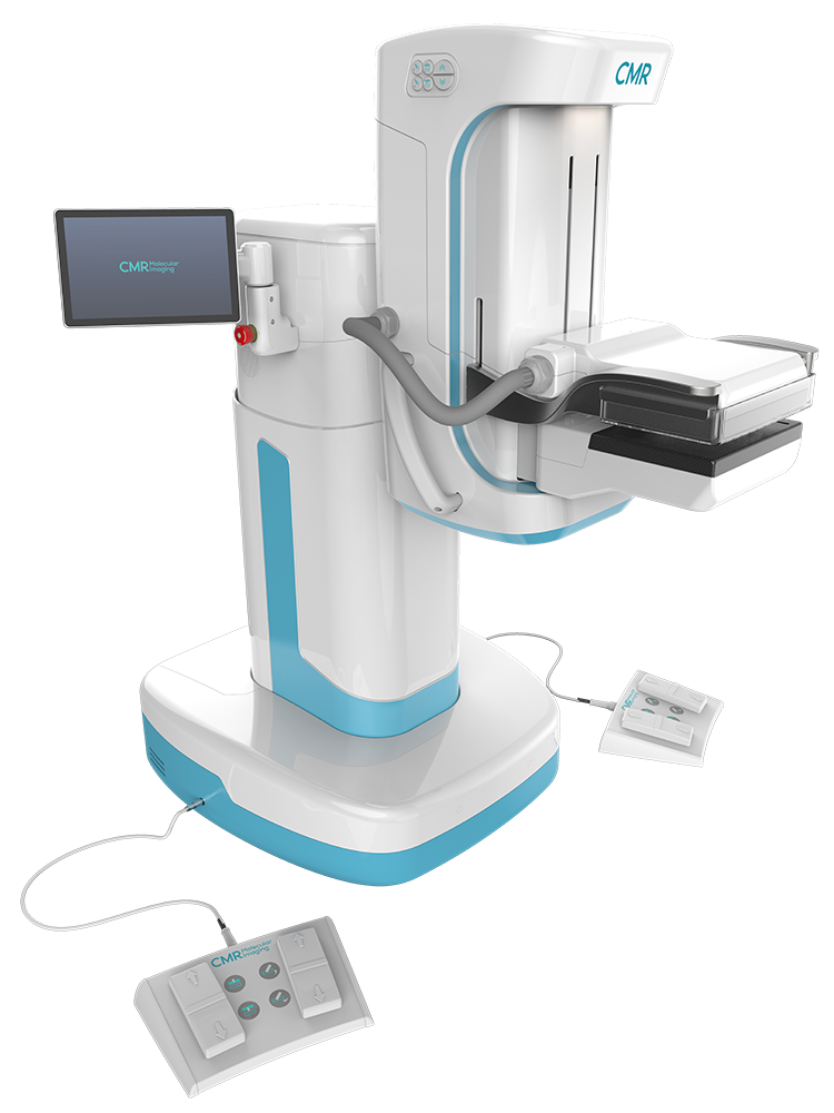
The LumaGEM® Molecular Breast Imaging system’s patented mounting mechanism allows the use of light breast immobilization for greater patient comfort during breast cancer imaging exam.
The LumaGEM MBI system for breast cancer imaging visually illuminates the enhanced metabolic activity in the breast associated with the occurrence of a tumor growth that is typically not visible on a digital mammogram due to tissue density and/or tumor size.
MBIs are extremely well tolerated by patients, as it’s a patient-friendly and a cost-effective tool for detecting hard to find small lesions in women who have dense breast tissue. When used as a secondary screening modality following digital mammography, the LumaGEM MBI system has increased breast cancer detection rates by almost 400% in early-stage invasive cancers.1
- If 1,000 women are screened with traditional digital mammography, 3 women would be found to have cancer.
- If 1,000 women were to be screened with digital mammography and MBI, nearly 12 women would be found to have cancer.1
The LumaGEM MBI system for breast cancer imaging is a functional imaging technology that measures the distribution of radionuclides by means of photon detection in order to aid in the evaluation of lesions in the breast tissue. The system features include:
- Clear resolution (high intrinsic spatial resolution, 1.6mm), the best available detector on the market
- Offers clinically high accuracy with Low false negatives (high sensitivity, 91%)2 and Low false positives (high specificity, 93%)2
- Knowing what is negative (Negative Predictive Value, 99.8%)2
- Proven/published lower dose capabilities minimizing patient risk (2.6mSv at 8 mCi)3,4
- Patient friendly, comfortable exam with minimal compression
- Alternative for women contraindicated for MRI due to pacemakers or other ferromagnetic implants, poor renal function, claustrophobia, and body size or gadolinium allergies
- Seamlessly integrates into workflow: Looks and acts like a mammography system with easy comparison to standard mammographic views (cranial caudal and mediolateral oblique)
The LumaGEM MBI system received FDA 510(k) clearance in 2011, and is installed in hospitals and breast imaging centers around the country.
What Separates MBI from the Past
Digitizing Molecular Breast Imaging: Earlier versions of this technology were introduced roughly 15 years ago. The LumaGEM represents a significant leap forward in technology and performance with fully digital, dual head camera detectors. With these improvements, LumaGEM customers have been able to identify cancers as small as 5mm in women with dense breast tissue, with greater than 90% sensitivity and specificity.
Awards and Recognition
- Bronze Award Winner in the Radiological and Electromechanical Devices Category of the 17th Annual Medical Design Excellence Awards (MDEA) competition
- Winner: Frost & Sullivan 2014 Best Practices Award – Global Breast Imaging Systems Product Leadership Award
LumaGEM Specifications At a Glance
LumaGEM is a planar, dual head, fully solid state digital imaging system utilizing cadmium zinc telluride (CZT) technology. LumaGEM delivers scans with a groundbreaking 1.6 mm intrinsic spatial resolution and 4.5% FWHM energy resolution.
| Detector Configuration | Dual head |
| Detectors | Cadmium Zinc Telluride (CZT) |
| Typical Clinical Dose1 | 8 mCi (99mTc-Sestamibi) |
| Whole-Body Dose2,3 | 2.4mSv (at 8 mCi) |
| Intrinsic Spatial Resolution | 1.6mm |
| Energy Resolution: Full-Width Half-Maximum (FWHM) | <5% (at 140 keV) |
| Sensitivity2 | 91% (with mammo) |
| Specificity2 | 93% |
| Collimator | Registered tungsten |
| Field-of-View | 16×20 cm2 |
- Rhodes DJ, Hruska CB, Conners AL, et al. JOURNAL CLUB: Molecular Breast Imaging at Reduced Radiation Dose for Supplemental Screening in Mammographically Dense Breasts. American Journal of Roentgenology. 2015;204(2):241-251.
- Rhodes DJ, Hruska CB Phillips SW, Whaley DH, O’Connor MK. Dedicated Dual Head Gamma Imaging for Breast Cancer Screening in Women with Mammographically Dense Breasts. Radiology. 2011; 258:106-118.
- Hendrick RE. Radiation Doses and Cancer Risk from Breast Imaging Studies. Radiology, 2010;257:246-253.
- O’Connor MK, Rhodes DJ, Hruska CB, Clancy CB, Vetter RJ. Comparison of Radiation Exposure and Associated Radiation-induced Cancer Risks from Mammography and Molecular Imaging of the Breast. Med Phys, 2010;37:6187-6198.