
A New Standard of Care in Early Breast Cancer Detection
Even when a mammogram is deemed negative, additional adjunctive tests, such as Molecular Breast Imaging (MBI), for women with dense breast tissue or who are considered high-risk for breast cancer should be considered.
When a patient is determined to have dense breast tissue, or has a suspicious mammogram, MBI offers a secondary screening option for breast cancer imaging that is more sensitive and specific. As a functional imaging modality, MBI provides the ability to have confidence in the diagnosis, and provide patients and physicians alike with peace of mind.
Since implementing MBI,…false positive rates have dropped significantly, and the cancers that are being caught with MBI are invasive, small and node-negative. We have been able to find cancers as small as 2-3mm in women with dense breast tissue, with over 90% sensitivity and specificity. Thankfully, the negative predictive values with MBI are extremely good, approaching 97% – 98%. This takes away much of the stress we were used to when diagnosing a patient. MBI really helps to determine what’s going on in women with a dense or complex mammogram. The results are easy to interpret and are incredibly helpful, which is why our breast surgeons love it. Many of them are even using MBI for preoperative surgical planning when MRI isn’t an option.
–Dr. Robin Shermis, Director of the ProMedica Breast Care Imaging Center, Toledo, Ohio
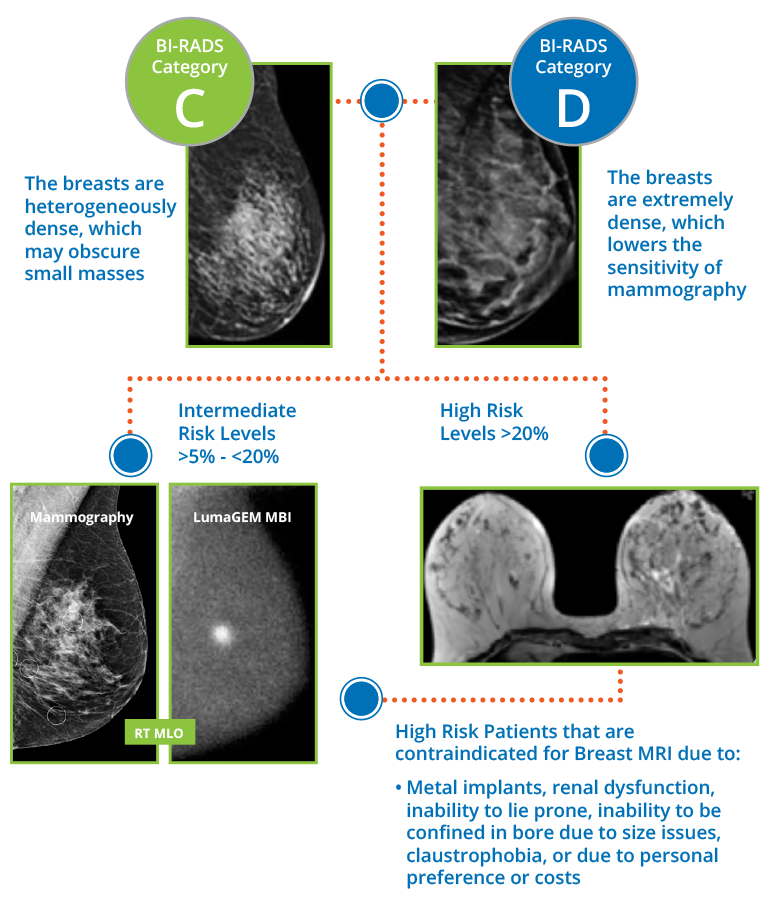
The Ideal MBI Workflow/Patient Algorithm
Molecular Breast Imaging is used as an adjunct/second look modality for dense breast women with lifetime risk of less than 20% after their annual mammograms. The images produced by an MBI exam are the same standard screening views as mammography, cranial caudal (CC) and mediolateral oblique (MLO), and are often read side by side with the patient’s prior mammogram. Users praise the fact that images are read in seconds and describe MBI as the “shortest path to the best care, at the lowest cost.”
With a demonstrated sensitivity and specificity over 90%, and 99.9%, negative predictive value, using MBI for dense breast cancer imaging delivers a strong value proposition to doctors by providing workflow efficiency, improved outcomes, and patient satisfaction.1
For the small minority of dense breast women whose risk factors place them at greater than 20% lifetime cancer risk (i.e., BRCA gene mutation, first degree relative with breast cancer, etc.), MRI is generally indicated. For those patients who have a 20% or greater lifetime risk, but are contraindicated and/or cannot tolerate an MRI, MBI also offers a solution for early breast cancer detection and imaging in patients with dense breast tissue.
The Ideal Molecular Breast Imaging (MBI) Workflow/Patient Algorithm
- Rhodes DJ, Hruska CB Phillips SW, Whaley DH, O’Connor MK. Dedicated Dual Head Gamma Imaging for Breast Cancer Screening in Women with Mammographically Dense Breasts. Radiology. 2011; 258:106-118.